Vitoria Olyntho
B.H.Sc. (Honours), Year 3
Please describe your involvement in any or all of your research work so far, including any achievements, awards, recognitions, or reflections. This can also include any work you have done to uplift and support the undergraduate research community, as well as details of any challenges you faced during your journey.
Growing up, I had an insatiable curiosity about the world, often pestering my teachers and family with hundreds of questions. My first memory in research was observing, in awe, hundreds of caterpillars hatch in my elementary school playground. I was intrigued and decided to look in the small "non-fiction" of my school's library to inquire about their life cycle. Into high school, I continued asking questions and seeking answers, always pursuing a deeper understanding of the world around me. As such, research became a passion of mine, and I was set on pursuing these interests further.My first research project was in my senior year of high school. Under the supervision of a lab technician at my high school, and in collaboration with the University of British Columbia, I cultured E. coli samples at diverse salt concentrations. My goal was to investigate bacterial resistance to high-solute environments. The research itself was simple: plating agar dishes, preparing nutrient-rich growth media, streaking bacteria onto the plates, and incubating them. However, under the non-sterile environment of my high school lab, many of the samples became contaminated and yielded inconclusive results. This was my first time experiencing failure in science (certainly not the last!). Every hurdle only motivated me more, and I was even more certain of my research aspirations. Having grown up in Brazil, I developed an interest in neglected tropical diseases (NTD) and wanted to explore them further. Dengue Fever (DF) is an NTD that was endemic in some regions in Brazil in 2016. My first encounter with Dengue was when I sat at my grandfather’s bedside as he fell ill with the disease. As someone who always carried an air of resilience, I was floored seeing my grandpa drenched in sweat, with barely enough energy to speak. I found that DF is a disease that is easily preventable. Since its virus is carried by a mosquito vector, its spread is greatly dependent on the availability of reproduction sites for mosquitoes. As such, awareness campaigns in Brazil focus on encouraging citizens to prevent still water to reduce reproduction sites. However, by the time awareness campaigns reach endemic regions, mosquitoes have often already replicated and viral transmission is at its peak. This prompted me to work on a project to investigate what factors contribute to the spread of Dengue and whether it would be possible to predict the spread of the disease to ensure timely awareness campaigns. Using a combination of mathematical modelling and data science, I developed a model that could predict the spread of Dengue based on climatological factors such as temperature and precipitation. Although this model was relatively simplistic, considering only a few variables involved in disease prevalence, it was able to predict the spread of Dengue two years in the future with some degree of success. In my first year at McMaster, in collaboration with two other undergraduate students, I authored an article proposing a protocol to evaluate the efficacy of three novel monoclonal antibody therapies for the treatment of leukemia. We submitted the abstract of this article in a competition by the Undergraduate Research in Natural and Clinical Science and Technology (URNCST) for the Mentored Journal program. Our project was selected and we worked under the supervision of a graduate student to draft a manuscript. After peer review, we published our article in the URNCST journal titled “Efficacy of Different Immunological Approaches Targeting CD22 for the Treatment of Relapsed or Refractory Acute Lymphoblastic Leukemia: A Research Protocol” (https://doi.org/10.26685/urncst.340). This project sparked in me an interest in immunology as I appreciated the complexities of the interactions and mechanisms studied in the field. I pursued these interests further during my second year of university, where I enrolled in a handful of immunology courses. I met Dr. Joshua Koenig during office hours for an introductory immunology course and soon joined him at the Jordana-Koenig Lab at the McMaster Immunology Research Centre. Under the supervision of Dr. Koenig, I undertook an immunofluorescence imaging project to characterize immune cells in allergic mouse tissues. This project was led by my mentor Jake Colautti, who taught me how to prepare tissues for imaging. The aim of this project was to investigate the intimate immune interactions required for the generation of immune responses in situ. Often, the challenge in characterizing immune cells is that they are incredibly diverse, something that cannot be captured adequately with traditional microscopy. To address this issue, a cyclic imaging technique named IBEX developed by one of our collaborators increases the multiplexity of tissues by inactivating fluorophores to allow for consecutive rounds of staining. This technology, however, has yet to be adapted to study allergic tissues. My project has been to optimize and validate antibodies that study markers relevant to allergy in mice for IBEX. In the summer, with support from the 2022 Bachelor of Health Sciences Summer Research Scholarship, my goal was to extend the multiplexity of an allergic mouse intestine to image 20 markers on the same tissue. These included structural and immune cell markers to characterize the immune microenvironment of the mouse intestine. During this time, a few confocal microscope demonstrations were happening at the McMaster Centre for Advanced Light Microscopy. I was tasked with evaluating how well these instruments could work to perform multiplexed immunofluorescence on our tissues, including their ability to image many fluorophores simultaneously. These demonstrations allowed me to develop my skills in microscopy, as well as work on my goal of visualizing 20 markers on an intestine sample. For one of the demonstrations, I was able to visualize ~19 markers in 4 rounds of imaging, successfully meeting my goal. However, having access to the microscope for an extra day of imaging, I decided to extend an extra round to reach ~24 markers: the most multiplexed image ever obtained at our lab. Up until this moment at the lab, I was always just learning new techniques and observing my mentors conduct experiments. However, it felt extremely rewarding to be able to apply my training to a project of my own and have an impact at the lab.
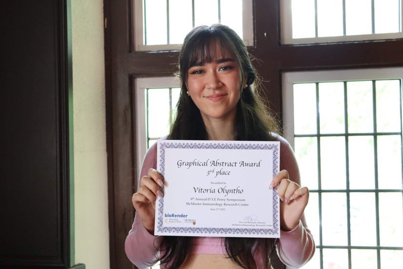
In June of this year, I shared our lab’s experiences with immunofluorescence imaging at the 8th Annual D.Y.E. Perey Symposium, hosted by the McMaster Immunology Research Centre, via a short speech. This event was the highlight of my summer. It felt incredible sharing my work with scientists at many levels of training and engaging in talks about immunology. This event also involved a Graphical Abstract competition sponsored by BioRender, where I was awarded 3rd place.
My work with Dr. Koenig has opened up many opportunities for me to collaborate with other scientists within McMaster and even internationally. In the past months, I developed my mentoring skills by teaching graduate students how to process and stain tissues, as well as designing antibody panels for multiplexed immunofluorescence. I am very grateful for these types of connections as I am both able to pass on the skills I have acquired and give back to McMaster’s scientific community, but also learn from students with more knowledge and experience than me.Currently, my research project has been to characterize immune cell interactions within the human nasal polyp. My work involves optimizing fluorescence antibodies to study the cells within these tissues. IBEX has never been conducted on human nasal polyps, and as a tissue our lab has not previously explored, I have faced many obstacles. I have had to battle with tons of non-specific antibody binding and autofluorescence of the tissue. Additionally, antibodies require multiple rounds of optimization to find the best dilution, which proves time-consuming and requires a lot of patience and resilience. Nonetheless, I am motivated to continue and contribute to the imaging community with antibodies validated for nasal polyps. The challenge of establishing this new platform has been very exciting to me, and I am looking forward to experiencing success in this new endeavour.Of which of your outlined achievements, work, or challenges overcome are you most proud?
Throughout my experiences in research, I have battled with feelings of insecurity and impostor syndrome. It often felt that my achievements had been a product of luck, or being at the right place at the right time. As such, I find myself discrediting much of my work, thinking anyone in my place would have been able to achieve what I did or more. However, one very seemingly insignificant, very simple experience made me feel like I belonged in research. This experience took place a few weeks ago when I was investigating nasal polyps, an endeavour that has proven very challenging. As described, this is partly because nasal polyps are extremely auto-fluorescent. I hypothesized that this was due to the presence of eosinophils in those tissues, which contain granules that fluoresce brightly. However, I had not yet confirmed the presence of eosinophils in the nasal polyp samples I imaged. Hence, I decided to run a round of immunofluorescence imaging with an eosinophil marker. My theory was that if the autofluorescence was co-expressed with the eosinophils, I could determine that they are, in fact, present in my samples. This would be a useful observation as I could then try strategies to prevent the fluorescence of their granules. I was preparing samples for imaging the following day. While preparing the samples, I thought to myself: what if the eosinophil stain does not work well? If the stain failed, the source of the autofluorescence would remain unclear, and it would be challenging to attempt to reduce it. My supervisor advised me to attempt a hematoxylin and eosin (H&E) stain of the nasal polyp sample, as eosinophils would appear bright with eosin. My biggest challenge was that our lab had only used H&E to stain blood smears in the past, and not tissues, which are processed very differently. As such, the protocol and materials available to me were specific to blood samples. I had four remaining unstained samples that I could use to optimize my H&E technique to visualize eosinophils.First, I searched for online resources on H&E protocols to follow. However, most of them required materials we did not have. One particular substance was a mounting medium that I searched the lab for, only to find that it had solidified after not being used for the past 10 years. I decided I had to improvise, troubleshoot, and optimize a protocol with the materials available. I used up two of my four tissues following an online tissue H&E protocol only to find that the hematoxylin I was using was staining too intensely. This protocol involved dipping the tissue in hematoxylin and eosin for a few seconds, so I decided to only dip the sample into hematoxylin for a fraction of a second to prevent over-staining. The stain initially appeared successful, but when visualizing the sample under the microscope, it was still too purple with hematoxylin.I had one sample left, and I felt ever so close to obtaining a proper stain. I decided to slightly dilute the hematoxylin and to only submerge the sample for a fraction of a second, immediately rinsing it after. After doing so, I was eager to finally look at the sample under the microscope. As soon as I focused the lens, I was ecstatic to see plenty of reddish granules across the nasal polyp. The granules I visualized seemed to belong to eosinophils. In fact, the tissue looked very similar to the images of nasal polyps I had identified in the literature.Truthfully, compared to high-end multiplexed immunofluorescence imaging, obtaining a simple H&E stain, which has been used by scientists since the 1800s, was nothing surprising or outstanding. However, looking through that microscope, I felt an immense sense of accomplishment and excitement. For the first time, I felt like a researcher, having successfully adapted a technique to achieve my goal, troubleshooting protocols along the way. This experience made me realize that science and discovery are based on trial-and-error, and how amazing it feels when you can finally experience success. There is much yet to come, as I have only just begun my journey in the field of research.


